Diagram Of Upper Leg Muscles And Tendons - Wiring And Diagram: Diagram Of Upper Leg Muscles And Tendons - Back muscles diagram 12 photos of the back muscles diagram back muscle workout diagram, back muscles diagram for massage, back muscles diagram massage, human back muscles diagram, upper back muscles.
Diagram Of Upper Leg Muscles And Tendons - Wiring And Diagram: Diagram Of Upper Leg Muscles And Tendons - Back muscles diagram 12 photos of the back muscles diagram back muscle workout diagram, back muscles diagram for massage, back muscles diagram massage, human back muscles diagram, upper back muscles.. Muscles of the leg include muscles of the thigh and foot. Many of the leg's muscles are also adapted to bipedalism, most substantially the gluteal muscles, the extensors of the knee joint, and the calf muscles.8. The anterior, lateral (fibular), superficial posterior, deep posterior compartments. Inferior surface of tarsals and metatarsals. Back muscles diagram 12 photos of the back muscles diagram back muscle workout diagram, back muscles diagram for massage, back muscles diagram massage, human back muscles diagram, upper back muscles.
A tendinous intersection is usually observed about the middle of the muscle. This is where the gto comes into play. Muscles in the arm diagram koibana info forearm anatomy upper limb anatomy arm muscle anatomy. Its tendon inserts on the dorsal surface of the base of 5th metatarsal bone. Tendons, fasciae and the various organs themselves depend on the muscular system and the functioning of muscle cells.
Calf Muscle Anatomy - Human Anatomy from www.verywellhealth.com Collectively, the muscles in this area in the lower part of the leg, the muscle belly combines with the soleus to from the calcaneal tendon, with inserts onto the calcaneus (the heel bone). Upper leg muscles and tendons. Related posts of muscle, tendons and ligaments of leg human. Lateral condyle and upper 2/3 of tibial shaft. This muscle originates on the distal anterior surface of the fibula and the adjacent interossous membrane. Its tendon inserts on the dorsal surface of the base of 5th metatarsal bone. Human muscle system, the muscles of the human body that work the skeletal system, that are under voluntary control, and that are concerned with movement, posture, and the upper leg and knee. Many of the leg's muscles are also adapted to bipedalism, most substantially the gluteal muscles, the extensors of the knee joint, and the calf muscles.8.
Muscles of the lower leg.
These changes negatively affect muscle quality, muscle and tendon stiffness and young's modulus and account for impairment in motor performance. Some are small in length, and others are thinner and less bulky than muscles that extend or flex the knee or primary superficial veins of right thigh and leg. Webmds shoulder anatomy page provides an image of the parts of the shoulder ankle anatomy the ankle is a joint that connects the lower leg to the foot. When studying the muscles of the leg, they can be compartmentalized into four primary groups: This is where the gto comes into play. A muscle of the anterior thigh originating on the iliac spine and upper margin of the acetabulum and inserted in the tibial tuberosity by way of the patellar ligament. Hand muscles and hand tendons. Related online courses on physioplus. Tendons, fasciae and the various organs themselves depend on the muscular system and the functioning of muscle cells. Each of these muscles is a discrete organ constructed of skeletal muscle tissue, blood vessels, tendons, and nerves. Muscles of the leg include muscles of the thigh and foot. The hamstring muscles are flexors, moving the upper leg (femur) at the hip joint and the lower leg (tibia and fibula). Causes of upper leg pain related to trauma may include the following.
Inferior surface of tarsals and metatarsals. The deep muscles that impact leg movement are generally smaller that those that are directly involved in flexing the knee. Tendons attach muscle to bone. These changes negatively affect muscle quality, muscle and tendon stiffness and young's modulus and account for impairment in motor performance. Between the tendons is a space called the popliteal fossa, with a small fat pad.
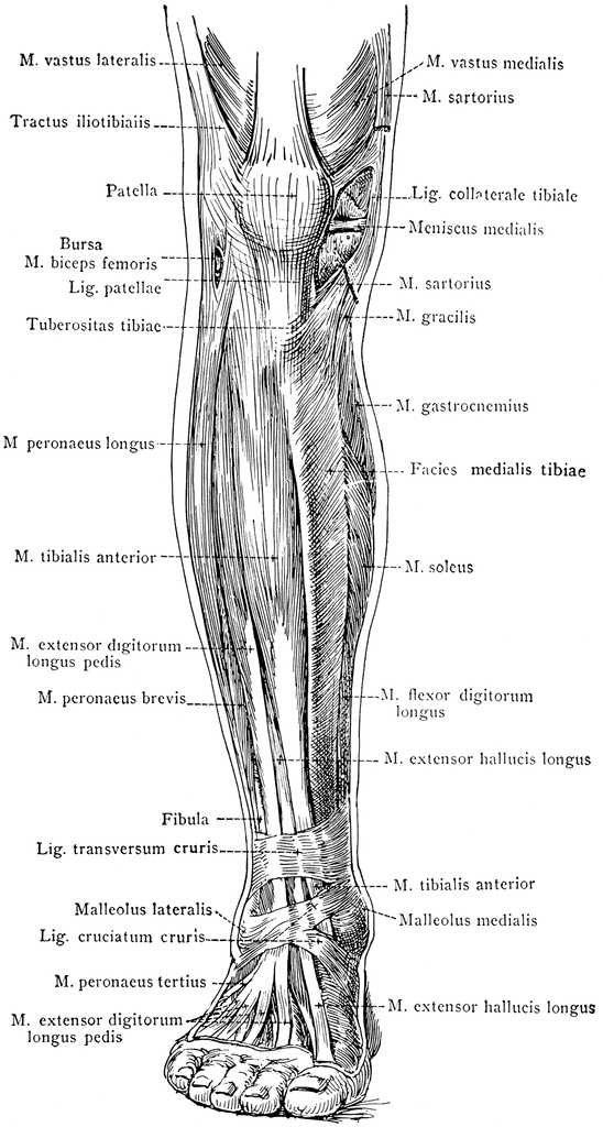
Anterior View of the Superficial Muscles of the Leg ... from etc.usf.edu Inferior surface of tarsals and metatarsals. When studying the muscles of the leg, they can be compartmentalized into four primary groups: Each of these muscles is a discrete organ constructed of skeletal muscle tissue, blood vessels, tendons, and nerves. Anatomy of leg and foot human muscular system. The anterior, lateral (fibular), superficial posterior, deep posterior compartments. Many of the leg's muscles are also adapted to bipedalism, most substantially the gluteal muscles, the extensors of the knee joint, and the calf muscles.8. This is where the gto comes into play. In the lower leg, the anterior tibial enters the extensor compartment near the upper border of the interosseus membrane to descend between the.
The accompanying muscle diagram further reveals the positions of the muscles in this pose.
Muscles of the lower leg. A muscle along the outside of the leg that bends the foot out at the ankle. Tendonitis is usually seen after excessive repetitive movement with which the tendon gradually becomes tighter until the fibers start to tear. Sartorius muscle appears from the anterior superior iliac spine and upper half of the notch immediately below it. Between the tendons is a space called the popliteal fossa, with a small fat pad. Some are small in length, and others are thinner and less bulky than muscles that extend or flex the knee or primary superficial veins of right thigh and leg. These changes negatively affect muscle quality, muscle and tendon stiffness and young's modulus and account for impairment in motor performance. The anterior, lateral (fibular), superficial posterior, deep posterior compartments. Muscles in the arm diagram koibana info forearm anatomy upper limb anatomy arm muscle anatomy. Foot muscles and tendons ã¢â?â? When studying the muscles of the leg, they can be compartmentalized into four primary groups: The upper part of the aponeurosis is curved backward over the upper edge of the tendon of the gracilis it is the great extensor muscle of the leg, forming a large fleshy mass which covers the front and sides of the femur. By striking in at a 90 degree angle into the bone, pain and dysfunction will.
Lateral condyle and upper 2/3 of tibial shaft. Related posts of muscle, tendons and ligaments of leg human. Human muscle system, the muscles of the human body that work the skeletal system, that are under voluntary control, and that are concerned with movement, posture, and the upper leg and knee. The biomechanical effects of stretching. Sartorius muscle appears from the anterior superior iliac spine and upper half of the notch immediately below it.
Physio Health from 4.bp.blogspot.com The muscles of the foot mainly customize and improve the actions of the long tendons and help fine movements of the toes. Related posts of muscle, tendons and ligaments of leg human. The diagram of lungs (диаграмма легких) since. A tendon is the end part of a muscle that attaches the muscle to the bone. Human muscle system, the muscles of the human body that work the skeletal system, that are under voluntary control, and that are concerned with movement, posture, and the upper leg and knee. Foot muscles and tendons ã¢â?â? The upper part of the aponeurosis is curved backward over the upper edge of the tendon of the gracilis it is the great extensor muscle of the leg, forming a large fleshy mass which covers the front and sides of the femur. Tendons, fasciae and the various organs themselves depend on the muscular system and the functioning of muscle cells.
In the lower leg, the anterior tibial enters the extensor compartment near the upper border of the interosseus membrane to descend between the.
Lateral condyle and upper 2/3 of tibial shaft. Causes of upper leg pain related to trauma may include the following. Back muscles diagram 12 photos of the back muscles diagram back muscle workout diagram, back muscles diagram for massage, back muscles diagram massage, human back muscles diagram, upper back muscles. The human body muscle anatomy body anatomy anatomy study muscular system bjorn borg human anatomy and physiology blood pressure remedies muscle building. Tendons arm wrist anatomy arm muscle anatomy anatomy and physiology. Related posts of muscle, tendons and ligaments of leg human. Its tendon inserts on the dorsal surface of the base of 5th metatarsal bone. Some are small in length, and others are thinner and less bulky than muscles that extend or flex the knee or primary superficial veins of right thigh and leg. The deep muscles that impact leg movement are generally smaller that those that are directly involved in flexing the knee. Proximal phalanx of hallicus and tendon of extensor digitorum longus. A tendinous intersection is usually observed about the middle of the muscle. The diagram of lungs (диаграмма легких) since. A muscle along the outside of the leg that bends the foot out at the ankle.

:max_bytes(150000):strip_icc()/plantaris-muscle-rupture-2549380-v2-764517a4508848dca33aab92f71c2182.png)


0 Komentar